A prion is thought to be an infectious agent that is comprised entirely of a propagated, mis-folded (rouge) protein.
The rogue prion protein has been implicated in a number of diseases in a variety of mammals, including bovine spongiform encephalopathy (BSE, also known as "mad cow disease") in cattle and Creutzfeldt-Jakob disease (CJD) in humans.
All hypothesized prion diseases affect the structure of the brain or other neural tissue, and all are currently untreatable and are always fatal.
PrPC refers to the normal cellular protein, which is found in a multitude of tissues, while PrPSC refers to the prion form of PrPC, and is responsible for clogging brain cells, and causing cells to die eventually.
Hypothetically, Prions infect and propagate by refolding converting normal molecules of the protein into the abnormally structured form.
All known prions induce the formation of tightly packed beta sheets. This altered structure is extremely stable and accumulates in infected tissue, causing cell death and tissue damage.
This stability means that prions are resistant to denaturation by chemical and physical agents, making disposal and containment of these particles difficult.
Mechanism of Prion Propagation
Prion Protein Structure
How Prions Arise
Prions Madcow mechanism
Viriods
Viroids are infectious agents composed of a single piece of circular single stranded RNA which has some double-stranded regions.
Because of their simplified structures both prions and viroids are sometimes called subviral particles.
Viroids mainly cause plant diseases but have recently been reported to cause a human disease (Hepatitis D)
Human diseases caused by Viroids
The only human disease known to be caused by a viroid is hepatitis D. For hepatitis D to occur there must be simultaneous infection of a cell with both the hepatitis B virus and the hepatitis D viroid (co-infection)
There is extensive sequence complementarity between the hepatitis D viroid RNA and human liver cell 7S RNA, a small cytoplasmic RNA that is a component of the signal recognition particle, the structure involved in the translocation of secretory and membrane-associated particles.
The hepatitis D viroid causes liver cell death via sequestering this 7S RNA and/or cleaving it.
Transmission
The hepatitis D viroid can only enter a human liver cell if it is enclosed in a capsid that contains a binding protein. It obtains this from the hepatitis B virus.
The viroid then enters the blood stream and can be transmitted via blood or serum transfusions.
Virusoids
Virusoids are circular single-stranded RNAs that are dependent on plant viruses(Viroids) for replication and encapsidation.
The genome of virusoids consist of several hundred nucleotides and only encodes structural proteins.
Virusoids are not considered as viruses but as subviral particles. Since they depend on helper viruses, they are classified as satellites
Done By: Alyssa Huang
Wednesday, January 28, 2009 5:02 PM
Virus Host interaction
1st Stage- Attachment
It is a specific binding between viral surface proteins and their receptors on the host cellular surface.
This specificity determines the host range of a virus.
Example, the Human Immunodeficiency Virus (HIV) attacks only human's immune cells (mainly T cells), because its surface protein, gp120, can interact with CD4 and chemokine receptors on the T cell's surface.
2nd Stage- Entry, Direct Cell Membrane Fusion
Viruses take advantage of these processes in a number of ways, after they have attached to the cell surface via binding to a specific receptor.
The simplest is DIRECT MEMBRANE FUSION, where the virion membrane fuses with the cell membrane, and the virion nucleoprotein complex is delivered into the cell cytoplasm directly.
This is generally a pH-independent process, and requires only that the membrane be fluid (ie: temperature in the physiological range), and generally that some divalent cations be present.
The entry process for HIV is shown in the graphic below.
3rd Stage-Uncoating
Uncoating is a process that viral capsid is degraded by viral enzymes or host enzymes.
4th Stage-Replication
Replication involves assembly of viral proteins and genetic materials produced in the host cell.
5th Stage- Assembly
Assembly of infectious virions is dependent on the action of: (a) an aspartyl protease encoded by the viral pol gene and responsible for cleavage of the gag and gag-pol precursors into mature proteins (1,2) and (b) cellular N-protein myristoyl transferase (NMT) which adds myristic acid to the N-terminus of gag, gag-pol and nef viral polyprotein precursors(3).
6th Stage- Release
Viruses may escape from the host cell by causing cell rupture (lysis).
Enveloped viruses such as HIV typically "bud" from the host cell. During the budding process, a virus acquires the phospholipid envelope containing the embedded viral glycoproteins.
Done by: Toh Wan Nee
Sunday, January 25, 2009 2:09 PM
Class VII
Class VII viruses are Double stranded DNA viruses that replicate though a single stranded RNA intermediate.
An example of a virus from class VII is hepadnaviridae.
Hepadnaviridae
Hepadnaviruses are a family of viruses which can cause liver infections in humans and animals.
There are two recognized genera:
• Genus Orthohepadnavirus; type species: Hepatitis B virus • Genus Avihepadnavirus; type species: Duck hepatitis B virus
Virology
Genome
Hepadnaviruses have very small genomes of partially double-stranded, partially single stranded circular DNA.
The genome consists of two uneven strands of DNA.
One has a negative-sense orientation, and the other, shorter, strand has a positive-sense orientation.
It has a endogeneous DNA which depends on DNA polymerase. It make use of overlapping reading frame (ORF) and has an RNA intermediate.
Replication
Hepadnaviruses replicate through an RNA intermediate (which they transcribe back into cDNA using reverse transcriptase).
The reverse transcriptase becomes covalently linked to a short 3- or 4-nucleotide primer.
Most hepadnaviruses will only replicate in specific hosts, and this makes experiments using in vitro methods very difficult.
The virus binds to specific receptors on cells and the core particle enters the cell cytoplasm.
This is then translocated to the nucleus, where the partially double stranded DNA is 'repaired' by the cell to form a complete circle of DNA.
It then undergoes transcription by the host cell RNA polymerase and the transcript is translated by host cell ribosomes.
New virus particles are formed, which acquire lipid from the endoplasmic reticulum of the host cell, and the genome is packaged within these particles, which then bud off from the cell.
Hepadnavirus-infected cells translate the protein known as the virus surface antigen many times until there is too much protein to coat the virions formed.
These proteins then aggregate to form rod shapes, and it is this antigen, known as the Australian or hepatitis B surface antigen, which is released from the cell and which leads to a very strong immune response from the host.
Pathogenesis
Hepadna virus can cause chronic or acute infection. Chronic or acute infection will on the age of infection. 90% of neonates and 50% of young children infected will have chronic infection whereas only 5% to 10% of Immuno-competent adults who were infected will develop chronic infection.
Clinical Features
Acute
Acute clinical features can be from subclinical to fulminant.
Acute symptoms:
• Loss of appetite • Nausea • Vomiting • Fever • Abdominal Pain • Jaundice
About 90% to 95% of acutely infected adults recover without squeal and about 5% to 10% of acutely infected adults become chronically infected.
Chronic
Patients who are chronically infected will be in a chronic carrier state. They are still potentially infectious but have not symptoms and no abnormalities on laboratory testing.
Others will have clinically apparent chronic hepatitis.
Some will go on to develop cirrosis, which is the hardening of the liver and finally, hepatocellular carcinoma.
Lab Diagnosis
Diagnosis of Hepatitis B virus infection generally made on the basis of serology.
For infected patients, there will be detectable serum hepatitis B surface antigen (HBsAg).
Hepatitis B envelop surface Antigen (HbeAg) is also detectable in acute infection which is characterised by a high rate of viral replication.
IgM antibodies against core antigen are also detectable in serum.
Subsequently, IgG antibodies against the core are produced.
As acute infection resolves, IgG antibodies against core antigen persist and IgM antibodies and HbsAg becomes undetectable.
Most people who have had acute infection that resolves continue to have IgG antibodies against core antigen for life
Acutely infected patients who do not clear Hepatitis B Virus continues to have serum HbsAg.
Diagnosis of Hepatitis B is confirmed and prognosis is assessed by liver autopsy.
Treatment
The treatment for hepatitis B includes a course of alpha interferon; this is a very expensive treatment which lasts for around 12-15 weeks and makes the patient very sick.
It seems that the interferon treatment aims at kick-starting the host immune response to clear the infection; it is not the drugs that clear the infection.
There are reverse transcriptase inhibitors available as treatment; these drugs target the virus replication strategy by (as the name suggests) inhibiting reverse transcription.
Hepatitis B infection can also be prevented by means of a recombinant Hepatitis B surface antigen vaccine.
Done by: Jeremy Lee
Friday, January 23, 2009 4:45 PM
Retroviridae
A retrovirus is a virus with an RNA genome that replicates by using a viral reverse transcriptase enzyme to transcribe its RNA into DNA in the host cell.
The DNA is then incorporated into the host's genome by an integrase enzyme.
The virus thereafter replicates as part of the host cell's DNA. Retroviruses are enveloped viruses that belong to the viral family Retroviridae.
Description of virus The virus itself stores its nucleic acid, in the form of a +mRNA genome and serves as a means of delivery of that genome into cells it targets as an obligate parasite, and constitutes the infection.
Once in the host's cell, the RNA strands undergo reverse transcription in the cytosol and are integrated into the host's genome, at which point the retroviral DNA is referred to as a provirus.
It is difficult to detect the virus until it has infected the host.
Virion Structure The main virion components are: • Envelope: lipid bilayer which obtained from host plasma membrane • RNA • Proteins: consisted of gag proteins, protease (PR), pol proteins and env proteins.
Gag proteins are major components of the viral capsid which are about 2000-4000 copies per virion. Protease, on one hand, is expressed differently in different viruses.
It functions in proteolytic cleavages during virion maturation to make mature gag and pol proteins. Pol proteins are responsible for synthesis of viral DNA and integration into host DNA after infection.
Finally, env proteins play role in association and entry of virion into the host cell.
Properties: It is a spherical virion and has a ribonucleoprotein in central nucleoid within icohedral capsid.
It has an envelope with glycoprotein peplomers and has 2 copies of linear (+) sense ssRNA.
The virus have a 3’ polyadenylated tail and 5’ cap and has the enzyme reverse transcriptase which reverse transcribe the viral RNA to DNA.
The virus will form long terminal repeats before provirus DNA is being inserted into host genome.
Gag, pol, env genes, some regulatory genes and some oncogenes are present in the virus.
Multiplication
When retroviruses have integrated their own genome into the germ line, their genome is passed on to a following generation.
While transcription was classically thought to only occur from DNA to RNA, reverse transcriptase transcribes RNA into DNA.
The term "retro" in retrovirus refers to this reversal (making DNA from RNA) of the central dogma of molecular biology.
These inserts are transcriped by host's enzymes into new RNA molecules which enter the cytosol. Next, some of these RNA molecules are translated into viral proteins. For example, the gag gene is translated into molecules of the capsid protein, the pol gene is transcribed into molecules of reverse transcriptase, and the env gene is translated into molecules of the envelope protein.
It is important to note that a retrovirus must "bring" its own reverse transcriptase in its capsid, otherwise it is unable to utilize the infected cell's enzymes to carry out the task, due to the unusual nature of producing DNA from RNA.
Because reverse transcription lacks the usual proofreading of DNA replication, a retrovirus mutates very often.
This enables the virus to grow resistant to antiviral pharmaceuticals quickly, and impedes the development of effective vaccines and inhibitors for the retrovirus.
Genes
Retrovirus genomes commonly contain three open reading frames that encode for proteins that can be found in the mature virus:
• group-specific antigen (gag) codes for core and structural proteins of the virus; • polymerase (pol) codes for reverse transcriptase, protease and integrase; and, • envelope (env) codes for the retroviral coat proteins.
Provirus
This DNA can be incorporated into host genome as a provirus that can be passed on to progeny cells. In this way some retroviruses can convert normal cells into cancer cells. Some provirus remains latent in the cell for a long period of time before it is activated by the change in cell environment.
Seven subfamilies
Subfamily: Orthoretrovirinae
Alpharetrovirus
Betaretrovirus
Gammaretrovirus
Deltaretrovirus
Epsilonretrovirus
Lentivirus
Subfamily: Spumaretrovirinae
Spumavirus
Lets look at Human Immunodeficiency Virus (HIV), a type of Lentivirus.
Transmission
HIV can be transmitted via sexual contact, through blood and blood products and secretion of other bodily fluids like semen and vaginal secretion.
A mother can pass HIV to a child via the placenta, mucosa and breast milk.
Primary infection
During the acute stages, flu-like symptoms, fever, skin rash and swollen lymph nodes may be observable.
Virulence factors include rate of replication, propensity of mutate and cytopathogenicity.
The host immune system can reisist infection via supression by CD8 T supressor cells and presence of cytotoxic T-lymphocytes.
Asymptomatic stage During asymptomatic stage, there is no apparent disease. But there will be a fall in CD4 T lymphocytes (primary target cells).
Possible signs includes fatigue, depression, weight loss and memory disorder.
Symptomatic stage
AIDS-related complex will occur. They are diseases not considered difinitive of AIDS but may be attributed to HIV infection. It is a indication of defects in cell-mediate immunity.
Opportunistic infections as a result of fall in CD4 T lymphocytes in AIDS patients.
Therapy
Non-specific therapeutic management is used to boost general health. This includes vitamins, mineral, antioxidants and others.
Specific therapeutic management involves in antiretroviral therapy like:
• Neucleoside reverse transcriptase inhibitors includes AZT (azidothymidine) and 3TC (lamivudine) • Non-nucleoside reverse transcriptase inhibitors includes Efavirenz and Nevirapine • Protease inhobitors include Indinavir and Ritonavir
The inefficiency of reverse transcriptase causes the virus to rapidly mutate. A combine therapy is used to combat resistance.
Immunomodulation include enhancement of immune system through treatment with potential drugs that are still under study.
Vaccines that are still under development.
Diseases
Diseases that can occur in AIDS patients includes Kaposi’s Sarcoma, Oral Hairy Leukoplakia and Candidiasis.
Done by: Jeremy Lee
Tuesday, January 20, 2009 8:51 PM
Flaviviridae It is a spherical enveloped virion with the size of 40-50nm. The inner core contains protein C and the enveloped is made up of glycoprotein peplomers (E).
It is single linear (+) ssRNA which is very infectious.
Dengue is one of the symptoms for flaviviridae. The most important arbovirus is present in the in it and it started from Southeast Asia to Americas to pacific to Africa.
There are 4 different class of Dengue. They are:
- Den-1 - Den-2 - Den-3 - Den-4
Class Den-2 shows the greatest antigenic and genotypic distance from the others. Is has a protective immunity after infection homotypic.
Dengue Life cycle
Symtoms for Dengue Fever
- Many infectious asymptomatic.
- Acute infectious resulting in fever, severe headache, retro-orbital pain, nausea and vomiting
- Severe muscle and bone pain
- Maculopapular rash just before recovery
Severe Dengue fever might lead to DENGUE HAEMORRAHGIC FEVER (DHF) /DENGUE SHOCK SYNDROME (DSS).
DHF/DSS
The properties of DHF/DSS are:
- Prior infection and age key factors - Seldom occurs in individuals above 15 years - Similar to yellow fever in biphasic nature:
Initial symptoms similar to DF followed by remission
Sudden deterioration of patent condition
- Severe prostration, hypotension, circulatory collapse, bleeding and shock - Bleeding
Injection and punction sites
Gastrointestinal bleeding
Petechiae in skin, mucous membranes (mouth)
There are also 4 different classes grading of DHF:
- Grade I: Fever with non-specific, constitutional symptoms and only haemorrhagic manifestations bring positive tourniquet test
- Grade II: As for grade I, but with specific haemorrhagic manifestations
- Grade III: Signs of circulatory failure or hypotension
- Grade IV: Profound shock with pulse and blood pressure undectable Pathogenesis of DHF/DSS
- Not well understood despite intensive study-2 theories
- Virulent strain theory:
Some strains more virulent than others
Molecular studies show variations in sequences amongst different strains, within serotypes Early evidence pointed to DEN-2.
- Antibody enhancement:
Main theory for DHF/DSS
Main Cell target of DEN: monocytes/ macrophages
Most cases of DHF/DSS had prior infection or infants below 1 year had maternal Ab
Monkey experiments showed similar enhancement
Cause
- Immune system overreacting - Sever Acute Respiratory Syndrome
By: Toh Wan Nee
Monday, January 19, 2009 12:07 PM
picornaviridae
-The largest virus family found on earth -it is a naked virus -Very infectious due to (+) RNA as genome information (it can be directly use as an mRNA for translation and replicate) (e.g. rhino virus)
• Why It cause infection throughout the year: àVirus is abundant in nasal discharge àInfected people go to crowded area àPeople sneeze without using tissue worse the hand contact to other people Rhino virus only infect upper respiratory tract No vaccination -our body can produce immune (IgG) toward it but is it only last for a few years -impossible to create vaccine due to: àFirstly, there is no vaccination àSecondly, there are over 100 types of rhino virus àLastly, our body can produce immune toward it (free), therefore, not worthy to create (if there is vaccine toward it)
Control • Avoid people that have cold • Sneeze into tissue • If you are infected, stay at home
Done by: Pow Ze Liang
Sunday, January 18, 2009 3:30 PM
orthomyxoviridae
-it is a enveloped virus - It have protein spikes on the envelope hemaggutinin(HA)/neuraminidase(NA)
- It have 8 segment of RNA as their genome information - Each segment have 3 polymerase - E.g. influenza A and B
• why cause infect through out the year due to: Contact with people that suffer from infection Virus is in abundant during nasal discharge Stress cause low in immune and infect easily
Influenza virus infect respiratory tract • 2 type of vaccine: Anti HA Ab IgA and IgG
Antigenic shift/drift • Antigen drift (occur in type A&B) Mutation in genetic code of the envelope antigen (HA/NA) • Antigen shift (occur in type A) Genes re-assortment
Strain naming convention • A/singapore/10/57 (H1N1) Influenza virus type A Occur in singapore Isolated 10 infect people It happen in year 1957 Subtype of the virus H1 and N1
Control • Vaccine against influenza but useless if antigen drift or shift occur • Antiviral drug like receptor analog and protease inhibitors
Done by: Pow Ze Liang
Thursday, January 15, 2009 1:16 PM
Class III
Class III viruses are double stranded RNA viruses that regulates replication, transcription, and translation separately, and has segmented genome. All replication activity is in cytoplasm, not nucleus.
An example of a virus from class III is Reoviridae.
Reoviridae
Reoviridae is a family of viruses that can affect the gastrointestinal system (such as Rotavirus) and respiratory tract.
Viruses in the family Reoviridae have genomes consisting of segmented, double-stranded RNA (dsRNA).
Reovirus infection occurs often in humans, but most cases are mild or subclinical.
The virus can be readily detected in feces, and may also be recovered from pharyngeal or nasal secretions, urine, cerebrospinal fluid, and blood.
Despite the ease of finding Reovirus in clinical specimens, their role in human disease or treatment is still uncertain.
Twelve genera of Reoviridae exist and are divided based on the presence of a "turret" protein on the inner capsid.
Turreted
Genus Aquareovirus: type species Aquareovirus A
Genus Cypovirus: type species Cypovirus 1 (CPV 1)
Genus Fijivirus: type species Fiji disease virus
Genus Idnoreovirus: type species Idnoreovirus 1
Genus Mycoreovirus: type species Mycoreovirus 1
Genus Orthoreovirus: type species Mammalian orthoreovirus
Genus Oryzavirus: type species Rice ragged stunt virus
Nonturreted
Genus Coltivirus: type species Colorado tick fever virus (CTFV)
Genus Orbivirus: type species Bluetongue virus
Genus Phytoreovirus: type species Rice dwarf virus
Genus Rotavirus: type species Rotavirus A - a common cause of primarily early childhood diarrhea
Genus Seadornavirus
Virology
Genome
The genome of rotavirus consists of 11 unique double helix molecules of RNA which are 18,555 nucleoside base-pairs in total.
Each helix, or segment, is a gene, numbered 1 to 11 by decreasing size. Each gene codes for one protein, except genes 9 and 11, which each code for two.
The RNA is surrounded by a three-layered icosahedral protein capsid. Viral particles are up to 76.5 nm in diameter and are not enveloped.
Replication
A simplified drawing of the rotavirus replication cycle
Rotavirus infects enterocytes of the villi of the small intestine, leading to structural and functional changes of the epithelium.
The triple protein coats make them resistant to the acidic pH of the stomach and the digestive enzymes in the gut.
The virus enter cells by receptor mediated endocytosis and form a vesicle known as an endosome.
Proteins in the third layer (VP7 and the VP4 spike) disrupt the membrane of the endosome, creating a difference in the calcium concentration.
This causes the breakdown of VP7 trimers into single protein subunits, leaving the VP2 and VP6 protein coats around the viral dsRNA, forming a double-layered particle (DLP).
The eleven dsRNA strands remain within the protection of the two protein shells and the viral RNA-dependent RNA polymerase creates mRNA transcripts of the double-stranded viral genome.
By remaining in the core, the viral RNA evades innate host immune responses called RNA interference that are triggered by the presence of double-stranded RNA.
During the infection, rotavirus produces mRNA for both protein biosynthesis and gene replication.
Most of the rotavirus proteins accumulate in viroplasm, where the RNA is replicated and the DLPs are assembled.
Viroplasm is formed around the cell nucleus as early as two hours after virus infection, and consists of viral factories thought to be made by two viral nonstructural proteins: NSP5 and NSP2.
Inhibition of NSP5 by RNA interference results in a sharp decrease in rotavirus replication.
The DLPs migrate to the endoplasmic reticulum where they obtain their third, outer layer (formed by VP7 and VP4).
The progeny viruses are released from the cell by lysis.
Pathogenesis Rotavirus gastroenteritis is a mild to severe disease characterised by vomiting, watery diarrhoea, and low-grade fever.
Once a child is infected by the virus, there is an incubation period of about two days before symptoms appear.
Symptoms often start with vomiting followed by four to eight days of profuse diarrhoea.
Dehydration is more common in rotavirus infection than in most of those caused by bacterial pathogens, and is the most common cause of death related to rotavirus infection.
Clinical Features
Acute
first infection usually produces symptoms, symptomatic infection rates are highest in children under two years of age severe symptoms tend to occur in children six months to two years of age, the elderly, and those with compromised or absent immune system functions
Chronic subsequent infections are typically asymptomatic,as immune system provides protection Infection in newborn children, although common, is often associated with mild or asymptomatic
disease
• Due to immunity acquired in childhood, most adults are not susceptible to rotavirus; gastroenteritis in adults usually has a cause other than rotavirus, but asymptomatic infections in adults may maintain the transmission of infection in the community.
Symptomatic reinfections are often due to a different rotavirus A serotype.
Lab Diagnosis
Specific diagnosis of infection with rotavirus A is made by identification of the virus in the patient's stool by enzyme immunoassay.
There are several licensed test kits on the market which are sensitive, specific and detect all serotypes of rotavirus A.
Other methods, electron microscopy and polyacrylamide gel electrophoresis, are used in research laboratories.
Reverse transcription-polymerase chain reaction (RT-PCR) can detect and identify all species and serotypes of human rotavirus.
Treatment and prognosis
• nonspecific • involves management of symptoms • maintenance of hydration. • oral rehydration with plain water, water plus salts, or water plus salts and sugar • Serious infections - hospitalisation where fluids are given by intravenous drip or nasogastric tube and child's electrolytes and blood sugar are monitored
Done by: Alyssa Huang
Tuesday, January 13, 2009 1:55 PM
Class II
Class II viruses are single stranded DNA viruses that replicate in the nucleus of the host cell to form a double stranded DNA. Single stranded DNA daughters come from these double stranded DNA.
An example of a virus from class II is Parvoviridae.
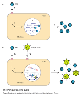
Parvoviridae
The Parvoviridae family includes the smallest known viruses, and some of the most environmentally resistant. They were discovered during the 1960s and affect vertebrates and insects.
The following genera are included here:
Subfamily Parvovirinae
• Genus Parvovirus; type species: Murine minute virus • Genus Erythrovirus; type species: B19 virus • Genus Dependovirus; type species: Adeno-associated virus 2 • Genus Amdovirus; type species: Aleutian mink disease virus • Genus Bocavirus; type species: Bovine parvovirus
Subfamily Densovirinae
• Genus Densovirus; type species: Junonia coenia densovirus • Genus Iteravirus; type species: Bombyx mori densovirus • Genus Brevidensovirus; type species: Aedes aegypti densovirus • Genus Pefudensovirus; type species: Periplanta fuliginosa densovirus
Parvovirus is a virus under the parvoviridae family, and it infects dogs. In puppies, infected puppies usually die within 24hours, while in adult dogs, the cases are mainly subclinical.
Virology
Genome
Parvovirus, commonly truncated to parvo, is a genus of the Parvoviridae family linear, non-segmented single stranded DNA viruses with an average genome size of 5 kbp. Parvoviruses are some of the smallest viruses found in nature. Some have been found as small as 23nm. Replication
1. Virus penetrates into the host cell. 2. Uncoating, and release of the viral genomic ssDNA into the nucleus. 3. The ssDNA is converted into dsDNA by cellular proteins. 4. viral mRNAs are transcribed when host cell enters S phase and translated to produce viral proteins. 5. Replication occurs through rolling-hairpin mechanism, whith NS1 nickase binding covalently to the 5’ genomic end. 6. Individual ssDNA genomes are excised from replication concatemers by a process called junction resolution. 7. These newly synthesized ssDNA can either a) be converted to dsDNA and serve as a template for transcription/replication b) be encapsidated to form new virions that can bud out of the host cell. Pathogenesis of Parvoviruses B19 Infection
B19 is associated with the following:-
1. Erythema infectiosum 2. Aplastic crisis in patients with chronic haemolytic anaemias. 3. Fetal loss in pregnancy 4. Persistent infection in immunocompromised patients
Clinical Features
Acute
• maculopapular rash • slapped cheek appearance • followed by joint involvement.
1. Erythema infectiosum Two thirds of the cases resolve by 2 weeks and the majority by 4 weeks.
2. Aplastic Crisis;-
• occurs in patients with chronic haemolytic anaemia • viral-like illness with fever and constitutional symptoms, • followed by the onset of fatigue and anaemia. • patient usually quickly recovers within a week, with the Hb recovering to normal levels. Such crises generally occur in children, and do not usually recur in an affected individual.
3. Infection in Pregnancy;- increased fetal loss
4. Persistent infection in immunocompromized patients - persistent infection resulting in severe anaemia in the immunocompromised
Diagnosis
• Serological tests
Aplastic crisis in a patient diagnosed by finding a reduction in Hb of >= 2g and a reticulocyte count of less than 0.2%
• Detection of Virus;- fetal blood samples or autopsy material where DNA-DNA hybridization and PCR can be carried out.
• Antibody Detection;- Counter-immunoelectrophoresis using B19 virus as antigen, detects antibody of all classes.
• Class specific Ab may be detected by ELISA or RIA.
Treatment and Prevention
• Administration of HNIG in cases of persistent B19 infection in the immunocompromised. • Symptomatic therapy for erythema infectiosum rarely necessary. • Cases of aplastic crisis require transfusion of erythocytes until satisfactory Hb level is obtained • susceptible patients with chronic haemolytic anaemias should be isolated from patients with B19 induced aplastic crisis • Pregnancy should be allowed to proceed and carefully monitored • At delivery examination of the cord blood for B19 IgM reveal whether the virus crossed the placenta.
Done by: Alyssa Huang
Sunday, January 11, 2009 9:07 PM
Class I - dsDNA
A virus classified in class I possesses double-stranded DNA genomes and do not go through RNA stage.
There are 2 different classes found in class I. Firstly, replication exclusively nuclear; very dependent on the host cell factors.
And secondly, replication in cytoplasm; viral genome contains all factors for genome replication and transcription.
An example of virus in class I is Poxviridae.
Poxviridae
Properties
Poxiviridae, which is the largest family, where viruses can infect both vertebrate and invertebrate animals. It can stay air borne for a long period of time and infect people.
Their virions (viral particles) are generally enveloped though the intracellular mature virions form of the virus, which contains different envelope and is infectious.
They vary in their shape depending upon the species but are generally shaped like a brick or as an oval form similarto a rounded brick.
The most common are vaccinia but Monkeypox infections are rising as seen in west and central frican rainforest countries.
Small pox have been eradicated.
Life cycle
1st – virus is bind to a receptor found on the host cell surface; the receptors are thought to be Glycosaminoglycands (GAGs).
2nd – The virus will enter the cell where is will starts to uncoat. Uncoating of virus is a 2 steps process.
First, the outer membrane is removed as the particle enters the cell. Second, the virus particle is uncoated further to release the core into the cytoplasm.
3rd – The pox viral genes are expressed in 2 phases.
These genes known as early genes will expressed first, encoding the non-structural protein, including proteins necessary for replication of the viral genome, and are expressed before the genome is replicated.
Late genes are expressed after the genome has been replicated and encode the structural proteins to make the virus particle.
4th – The assembly of the virus particle occurs in the cytoskeleton of the cell and is a complex process. The replication of this virus is unusual for a virus with double stranded DNA genome because it encodes its own machinery for genome replication and therefore the replication occurs in the cytoplasm.
Most viruses with a double stranded DNA genome replicate in the nucleus and use the host cells genome replication machinery.
5th – This is the last stage of the cycle where the virus is being released to infect other surrounding cells.
Clinical Features
- Only infect human - Respiratory Secretions-Saliva - Always associated with skin lesions - At least 9 poxviruses cause disease in human - 7-12 days incubation - Initially influenza-like symptoms - Scarring of skins, blindness, DEATH
Lab Diagnosis
- Declared by WHO to be eradicated in 1980 - Not infectious during incubation but during symptoms - Aerosol or air droplets - BEST DEFENCE is VACCINATION
Control
- All infected materials must be incinerated - Last outbreak in Yugoslavia in 1972 due to accidental release - Vaccination stopped more than 30 years ago
By: Toh Wan Nee
Friday, January 9, 2009 12:23 PM
Herpesviridae
Herpesviridae is a large family of DNA viruses that cause diseases in animals and human.
It is an enveloped DNA virus and it can cause latent or lytic infections. It has large double-stranded, linear DNA genomes encased within an icosahedral protein cage called the capsid which is itself wrapped in a lipid bilayer membrane called the envelope. This whole particle is known as the virion.
There is no vaccine is currently available to prevent or eliminate herpes. However, treatments are available to reduce viral reproduction and shedding, prevent the virus from entering the skin, and alleviate the severity of symptomatic episodes.
There are 3 different group of Herpesviridae, they are classified as:
Herpes Simplex viruses are classified into 2 groups; Herpes Simplex virus 1 and Herpes Simplex virus 2. They enter and hide in the human nervous system, accounting for their durability in the human body. HSV-1 is commonly associated with herpes outbreaks of the face known as cold sores or fever blisters, whereas HSV-2 is more often associated with genital herpes.
Varicella Zoster virus
Varicella Zoster virus (Human Herpes 3) are also classified into 2 groups; Varicella which is chicken pox-respiratory tract and the other group is Herpes zoster is Shingles. Shingles is found in the second stage of chicken pox and it will occur many years after the initial infection.
Girl infected with chicken pox
Guy infected with shingles
Betaherpes: Cytomegalovirus, Human herpes type 6, Human Herpes type 7
Cytomegalovirus (Human herpes 5) infections are frequently associated with salivary glands, though they may be found throughout the body. HCMV infection can also be life threatening for patients who are immunocompromised (e.g. patients with HIV, organ transplant recipients). This virus can also be found in urine, semen, cervial secretion and even breast milk. It is also a virus most frequently transmitted to a developing fetus.
Gammaherpes: Epstein Barr Virus, Human herpes type 8
Epstein Barr Virus (Human Herpes 4) is one of the most common viruses in humans. Most people become infected with EBV, which is often asymptomatic but commonly causes infectious mononucleosis. It has surface receptors known as glycoprotein H (gH) which is essential for penetration of B cells but it also plays a role in attachment of virus to epithelial cells.
Life Cycle of Herpesviridae
(A) Herpes simplex virus (HSV) is shown undergoing the lytic cycle (entry, uncoating, and viral transcription and DNA replication in the nucleus, particle assembly, and exit from the cell) in epithelial cells of the skin to cause a primary infection.
(B) Some virus enters the sensory neuron terminals and travels retrogradely to the nucleus where it establishes latency.
(C) Periodic reactivation results in anterograde transport of viral particles, shedding from the neuron, and re-infection of epithelial cells, which leads to asymptomatic shedding or recurrent lesions.
Clinical Features of Varicella Zoster
Primary infection
- Fever - Lesion all over the body - Scratched lesion which will lead to secondary infection
Secondary Infection - Scarring - Dangerous in Pregnant women-unborn child will born limbless - May affect the nervous system- Guillain Barre syndrome
- Stop Risky Behaviour-mainly by sexual contact - Don’t kiss
By: Toh Wan Nee
Tuesday, January 6, 2009 11:31 AM
Baltimore classification
The Baltimore classification is a virus classification system which groups viruses into families depending on their type of genome (DNA, RNA, single-stranded (ss), double-stranded (ds) etc.) and their method of replication.
Classifications
Classifying viruses according to their genome means that those in a given category will all behave in much the same way, which offers some indication of how to proceed with further research. In short:
This type of virus usually must enter the host nucleus before it is able to replicate. Furthermore, these viruses require host cell polymerases to replicate the viral genome and hence are highly dependent on the cell cycle. Proper infection and production of progeny requires that the cell be in replication as that is when the cell's polymerases are active. The virus may induce the cell to forcefully undergo cell division, and chronically, this may lead to transformation of the cell and ultimately, cancer. Examples include Herpesviridae, Adenoviridae and Papovaviridae. There is only one well studied example in which a class 1 virus is not replicating within the nucleus, that is the Poxvirus family, a highly pathogenic virus that infects vertebrates and includes the smallpox virus.
Class II: Single stranded DNA viruses
Viruses that fall under this category include ones that are not as well studied, but still pertain highly to vertebrates. Two examples include the Circoviridae and Parvoviridae. They replicate within the nucleus and form a double stranded DNA intermediate during replication. A prevalent but asymptomatic human Circovirus called Transfusion Transmitted Virus (TTV) is included within this classification.
Class III: Double stranded RNA viruses
As with most RNA viruses, this class replicates in the cytoplasm, not having to use the host replication polymerases to as much a degree as DNA viruses. This family is also not as well studied as the rest and includes 2 major families, the Reoviridae and Birnaviridae. Replication is monocistronic and includes individual, segmented genomes, meaning that each of the genes code for only one protein, unlike other viruses which exhibit more complex translation.
Class IV & V: Single stranded RNA viruses
These viruses consist of two types, however both share the fact that replication is primarily in the cytoplasm, and that replication is not as dependent on the cell cycle as other DNA viruses. This class of viruses is one of the best studied, alongside the double stranded DNA viruses.
Class IV: Single stranded RNA viruses - Positive (+) sense The positive sense RNA viruses and indeed all genes defined as positive sense can be directly accessed by host polymerases to immediately form proteins. These can be divided into two groups, both of which reproduce in the cytoplasm:
• Viruses with polycistronic mRNA where the genome RNA forms the mRNA and is translated into a polyprotein product that is subsequently cleaved to form the mature proteins. This means that the gene can utilize a few methods in which to produce proteins from the same strand of RNA, all in the sake of reducing the size of its gene.
• Viruses with complex transcription, for which subgenomic mRNAs, ribosomal frameshifting and proteolytic processing of polyproteins may be used. All of which are different mechanisms with which to produce proteins from the same strand of RNA.
Examples of this class include the families Astroviridae, Caliciviridae, Coronaviridae, Flaviviridae, Picornaviridae, Arteriviridae and Togaviridae.
Class V: Single stranded RNA viruses - Negative (-) sense
The negative sense RNA viruses and indeed all genes defined as negative sense cannot be directly accessed by host polymerases to immediately form proteins. Instead, they must be transcripted by viral polymerases into a "readable" form, which is the positive sense reciprocal. These can also be divided into two groups:
• Viruses containing non segmented genomes for which the first step in replication is transcription from the (-) stranded genome by the viral RNA-dependent RNA polymerase to yield monocistronic mRNAs that code for the various viral proteins. A (+) sense genome copy is then produced that serves as template for production of the (-) strand genome. Replication is within the cytoplasm.
• Viruses with segmented genomes for which replication occurs in the nucleus and for which the viral RNA-dependent RNA polymerase produces monocistronic mRNAs from each genome segment. The largest difference between the two is the location of replication. Examples in this class include the families Arenaviridae, Orthomyxoviridae, Paramyxoviridae, Bunyaviridae, Filoviridae and Rhabdoviridae (the latter which includes rabies).
Class VI: Positive (+) sense single stranded RNA viruses that replicate through a DNA intermediate A well studied family of this class of viruses include the retroviruses. One defining feature is the use of reverse transcriptase to convert the positive sense RNA into DNA. Instead of using the RNA for templates of proteins, they use DNA to create the templates, which is spliced into the host genome using integrase. Replication can then commence with the help of the host cell's polymerases. A well studied example includes HIV.
Class VII: Double stranded DNA viruses that replicate though a single stranded RNA intermediate
This small group of viruses, exemplified by the Hepatitis B virus (which is in the Hepadnaviridae family), have a double-stranded, gapped genome that is subsequently filled in to form a covalently closed circle (ccc DNA) that serves as a template for production of viral mRNAs and a subgenomic RNA. The pregenome RNA serves as template for the viral reverse transcriptase and for production of the DNA genome.
Done by: Nicolina Ng
Sunday, January 4, 2009 3:01 PM
Introduction to viruses
Viruses
A virus is a biological agent that reproduces inside the cells of living hosts.
When infected by a virus, a host cell is forced to produce many thousands of identical copies of the original virus, at an extraordinary rate.
Unlike most living things, viruses do not have cells that divide; new viruses are assembled in the infected host cell.
All viruses have genes made from either DNA or RNA, long molecules that carry the genetic information; all have a protein coat that protects these genes; and some have an envelope of fat that surrounds them when they are not within a cell.
Viruses vary in shape from the simple helical and icosahedral to more complex structures.
The origins of viruses is unclear: some may have evolved from plasmids—pieces of DNA that can move between cells—while others may have evolved from bacteria.
Viral infections often cause disease in humans and animals, however they are usually eliminated by the immune system, conferring lifetime immunity to the host for that virus.
Antibiotics have no effect on viruses, but antiviral drugs have been developed to treat life-threatening infections.
Vaccines that produce lifelong immunity can prevent some viral infections.
Done by: Nicolina Ng
Friday, January 2, 2009 3:33 PM
SARS-CoV (Severe Acute Respiratory Syndrome-coronavirus) Do you know that? -it is a group IV virus (+)ssRNA -falls under the family of Coronaviridae -it is air borne transmit -it is an emerging virus
Introduction: In Singapore, the virus was introduced through 3 ladies that went to Hong Kong for tourism purpose. Under the same hotel, there is a professor from China is infected with SARS-CoV. The virus was spread through when the 3 ladies shared the same lift with him. Initially, there was no symptom. Till when came back to Singapore, there was not much symptom but fever and cough. After went to clinic. The symptoms still persist. They were admitted to the hospital and diagnosis that they were suffered from SARS-CoV. History of virus: The origin of SARS is still uncertain as not much research is done. Unique feature: -it is (+) RNA virus in Group IV. It is very infectious like rhino virus -it is a envelop virus -it is very sensitive to heat -it is unstable at ≥30̊C Where does the virus infect? -respiratory tract
Do you know the symptom? All symptoms will appear after 2-10 days of infection. Initial symptom -fever (≥38̊C) -myalgia -lethargy -cough -sore throat Late symptom -shortness of breath
Lab diagnosis: -PCR -immunofluorescence -ELISA
Treatment: - Vaccination (Prednisone) But negative effect is the immune response will decrease -since the virus is unstable with heat ≥30̊C, therefore, for now the virus will hardly infect any human.
How it replicate?
1. Attachment: the virus antigen is attaching the receptor of the host cell. In order to gain entry to the host cell (endocytosis) 2. Entry: the virus enter the cell (but till now still not clear that whether the virus fuse with the host cell membrane) 3. Uncoating: the virus uncoat it’s envelop allow gene expression to take place 4. Replication: The immediate genes produce RNA polymerase and start to duplicate its genome (early gene) the RNA of the virus produce (-) RNA and use it as a template to produce more (+) RNA. The late gene of the RNA will produce the protein that is needed to infect other cell but at a later time. 5. Assembly: the duplicated RNA will assemble with the protein that is produced to form virus 6. Release: once the virus is ready and it is release through exocytosis
Thursday, January 1, 2009 4:20 PM
WELCOME!!!
welcome to our humble blog. We have created this blog due to a module assignment given to us by our school.
In this blog, you will be learning about virology.
topics that we will be covering are :
Introduction to Virology
Baltimore Classification
Hepadnaviridae
Retroviridae
Othomyxoviridae
Picornaviridae
Poxviridae
Herpesviridae
Flaviviridae
Reoviridae
Parvoviridae
Virus Host Interaction
Prions, Viriods and Virisoids
Hope that after going through our blog, you knowledge on viruses will increase.
And now... let me introduce you the team of editors:
Left to the right: Jeremy Lee, Pow Ze Liang, Toh Wan Nee, Nicolina Ng and Alyssa huang
These are the 5 people who have worked very hard to present you this blog. So I hope that all of you will enjoy your stay here.
And if there is any comments about this blog or just a shout out... don't hesitate to use the tag box. Hope that all of you will leave a comment.
Thanks
By: Jeremy Lee
Us
Group Members:
Alyssa Huang
Jeremy Lee
Nicolina Ng
Pow Ze Liang
Toh Wan Nee
Module Group:
MB0803

 Mechanism of Prion Propagation


%5B1%5D.jpg)


